Launching 1st March 2023. Also check out: https://www.thailandmedical.news/
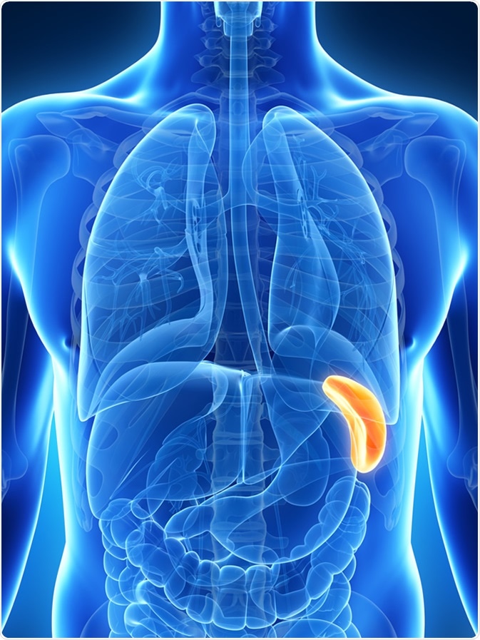
The spleen is an organ situated behind the stomach, in the upper left part of the abdomen known as the left hypochondrium. It is located under the ribcage and therefore is not usually felt by the palpating finger. However, in abnormal conditions it may give rise to pain felt as an aching sensation deep behind the left ribcage. This area may also hurt when it is touched. This could signal splenic damage, splenic rupture, or splenomegaly. Severe pain arising from the spleen often signals the presence of splenic infarction.

Damage to the spleen is rare because of its protected position under the left side of the ribcage. In spite of this, it may occur with a very forceful blow to the abdominal area, as may happen during a fight, a motor accident, during a contact sport, or because of a rib fracture, with the broken end of the rib penetrating the spleen. This may cause immediate rupture of the spleen, or it may result in the formation of a hematoma. This may later resolve partially to produce a splenic cyst. In other cases, splenic rupture may not actually occur for some weeks following the trauma.
Some signs of splenic rupture include:
Splenic rupture is always a medical emergency because it may be catastrophic in the degree of hemorrhage that occurs, and needs immediately evaluation and treatment.
Spleen enlargement, or splenomegaly, is another reason for splenic pain. This may occur due to a host of causes, whether traumatic, malignant, metabolic, or obstructive.
Causes of splenomegaly include:
Many drugs induce spleen pain by their adverse effects. These include:
The drug-induced adverse reactions are usually temporary and the spleen pain subsides with the spleen size once the drugs responsible are stopped.
Diagnosis of the cause of splenic pain is based upon the assessment of splenic size, hemodynamic parameters, history of injury or other medical conditions, and other symptoms. Palpation of the lower border of the spleen will reveal whether it is enlarged, because this occurs only after significant splenomegaly has occurred.
Splenic pain is evaluated using imaging with ultrasonic transducers or CT/MRI scanners. Treatment is directed at the underlying cause of the pain. Importantly, all contact with the organ should be avoided for a few weeks to avoid worsening the damage.