Launching 1st March 2023. Also check out: https://www.thailandmedical.news/
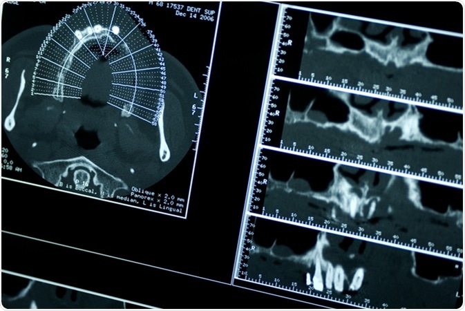
Microcomputed tomography (Micro CT) is a high-resolution imaging device, which produces non-destructive 3D images of a biological structure, like a tooth.

Micro CT can be compared to CT scan to visualize images of various structures in the body, but micro CT produces finer images with an ultra-high resolution on a minute scale.
Micro CT has varied uses in dentistry ranging from dental research to treatment. Micro CT produce high-quality images from the outer to innermost structure of the tooth and surrounding structures. As micro CT creates non-destructive images, the same image can be viewed multiple times and the image remains incessantly for biological and mechanical testing.
Enamel is the hardest and outmost structure of the tooth. Measuring thickness of enamel helps to understand various stages of human evolution. The older analysis technique was destructive and involved cutting the cross-sections of rare fossil specimens. However, sectioning the tooth of such specimens was objected to and remained a controversial topic among scientists.
Tooth CT scan also produced scattered and poor-quality images which hampered detailed examination. Micro CT has an edge over both the above-mentioned methods as it produces high-resolution and durable images of the enamel, leading to a precise measurement of enamel thickness. Besides this Micro CT can also accurately measure the width of inner tooth structures like dentin and pulp chamber.
Root canal treatment (RCT) is a procedure in which the infected tooth canals are properly cleaned and filled with an inert biocompatible tooth material. By leveraging Micro CT, an endodontist (dentist specialized to perform root canal treatment) can then access and evaluate root canal more effectively. Below are some uses of Micro CT during RCT:
It is vital to understand the morphology of the root canal during RCT. Some canals are tortuous or curved in a way that it is challenging for a dentist to obturate (filling with the inert filling material) during root canal treatment. Conventional CT can only produce 2D images which makes it difficult to analyze the root canal morphology. Whereas, Micro CT produces high-resolution 3D images which allow precise assessment of the root canals.
Micro CT has proved exceptional benefits in understanding the highly complex C-shaped canals. These root canals are usually found in molars of the mandible (lower jaw). Micro CT helps in analyzing the in-depth morphology of such canals during root canal treatment.
Root canal preparation is the most vital step during RCT. Root canal preparation determines the removal of infection from the root canal, proper creation of the space for infusing medications and preparing the canal for properly placing the filling material. Micro CT has an edge over widely used radiographs as it makes easier to determine the various parameters such as surface area and volume of the root canal.
Micro CT is also helpful in the accurate utilization of root canal instruments like pro taper files and hand K-files. These tiny needle-shaped instruments are used to take out root fillings in case of RCT failure and subsequent infection.
Currently, the main utilization of Micro CT is the analysis of trabecular bone (porous internal tissue of the skeletal bone). Micro CT is beneficial in understanding the growth and development of craniofacial bone. Micro CT has made easier to determine various parameters such as:
Micro CT has proved to be the revolutionary as it helps in understanding and analyzing the bone architecture and its growth and mineralization in a more effective way. It can also help assess craniofacial shape and abnormal growth.
FEM has recently gained importance in understanding the physical phenomenon in structural, solid, fluid mechanics, and biomechanics. Micro-CT helps to understand FEM of teeth and bones. Micro CT has enabled proper sectioning of enamel, dentin, and pulp into different parts based on parameters like pixel grey level values or mineral density, which allows understanding the of the amount of stress and strain required during cavity preparation and filling a carious tooth with a restorative material.
Tissue engineering aims to create bio-artificial organs in the laboratory. Tissue engineering is beneficial in providing permanent cure to individuals who have had severe trauma, birth defects (e.g., cleft palate), or terminal disease like cancer. Micro CT is beneficial in studying various stages of tissue engineering.
For a dental implant to be successfully fixed in the bone it is essential to determine the implant stabilization and osseointegration (connection between the living bone and the artificially placed implant). Micro-CT enables quick and high-resolution images which aid in assessing the connection of trabecular and cortical bone and the implant. Micro CT allows a clear visibility of bone growth formation over the implant surface which clearly determines the osseointegration and overall success of the implant.