Launching 1st March 2023. Also check out: https://www.thailandmedical.news/
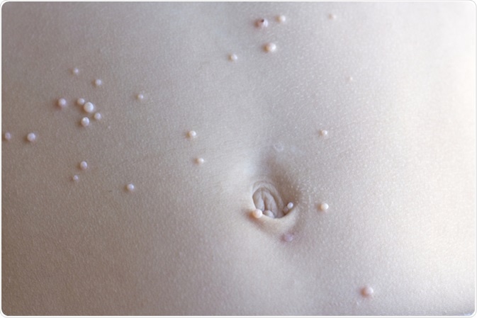
Molluscum contagiosum represents a superficial viral infection of the skin that is most commonly seen in children and young adults. Caused by the Molluscum contagiosum virus, this disease typically presents as smooth, small, flesh-colored and dome-shaped protrusions on the skin with a central indentation (umbilication).
After the eradication of smallpox, the molluscum contagiosum virus remains the only representative of the pox family of viruses to infect humans. However, unlike smallpox, infections with this virus were not clinically significant until the human immunodeficiency virus (HIV) stepped onto the stage.

The condition was named in 1817 by a British physician Thomas Bateman who observed the characteristic appearance of the lesions from which he managed to express a milky fluid. Molluscum contagiosum was not the only term used to describe this disease back then – phrases such as “molluscum epitheliale”, “molluscum verrucosum”, and even “acne varioliformis” have been observed in the literature.
But the Journal of the American Medical Association (JAMA) published a position paper in 1899 that cemented Batemen’s original name, and which also cited all the acquired evidence supporting the infectious nature of the papules. The viral nature was initially hypothesized by Julius Burg in 1905, whereas in 1933 it was already known that the infection is caused by a double-stranded DNA virus from the pox family.
In 1941, scientists Henderson and Paterson demonstrated specific intracytoplasmic viral inclusions that today bear their names (i.e. Henderson-Peterson or molluscum bodies). A structure known as Borrel bodies (akin to molluscum bodies) can also be seen in fowlpox-infected tissues.
As already mentioned, molluscum contagiosum virus is a linear, non-segmented, double-stranded DNA virus from the genus Molluscipoxvirus, subfamily Chordopoxvirinae and family Poxviridae. As the only species in the genus, molluscum contagiosum virus forms a group by itself that is separate from variola and vaccinia viruses, fowlpox virus, and all the other genera.
By employing DNA fingerprinting techniques, four characteristic genetic subtypes of molluscum contagiosum virus can be differentiated (MCV I, II, III and IV). A majority of human infections (more than 96%) are due to MCV I, albeit most infections (approximately 60%) in HIV-infected individuals are attributed to MCV II.
Morphologically, particles of molluscum contagiosum virus are pleomorphic (i.e. varying in shape and size), most often showing an ovoid or brick-shaped appearance. They possess an envelope, a surface membrane, and a central core that is dumbbell-shaped, as well as lateral bodies.
After direct skin contact with an infected individual, viral entrance is facilitated by superficial damage to the epithelium. This is followed by an abnormal proliferation of epithelial cells in the basal layer with subsequent acanthosis, leading to folded and hyperplastic epithelium.
The molluscum contagiosum virus encodes specific molecules that are used to control host defenses by utilizing several different methods to evade detection by the human immune system. Such viral evasion is responsible for the viral localization in the epidermal keratinocytes, the absence of inflammatory infiltrate and the lack of activation of specific T and natural killer cells of the immune system.
Better insights in the exact pathogenesis of molluscum contagiosum have been hindered by the lack of ability to replicate the virus in tissue cultures (due to a defect in the genome expression), as well as the absence of appropriate animal models. Although researchers have succeeded in infecting human skin with the virus and grafting it onto athymic mice, they have not been able to ensure continuing viral replication.